42 heart structure and labels
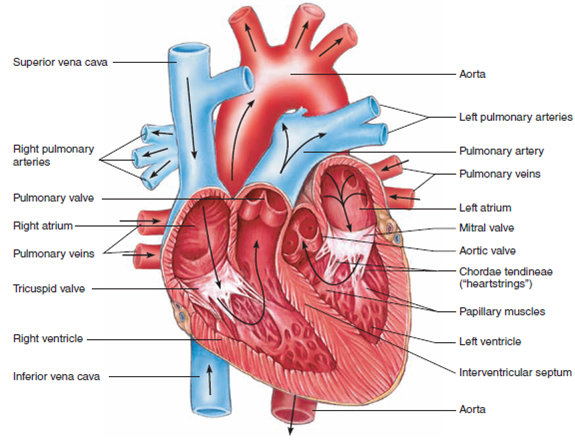
Structure Of The Heart | A-Level Biology Revision Notes The heart is a hollow muscular organ that lies in the middle of the chest cavity. It is enclosed in the pericardium, which protects the heart and facilitates its pumping action. The heart is divided into four chambers: The two atria (auricles): these are the upper two chambers. They have thin walls which receive blood from veins. Layers of the heart: Epicardium, myocardium, endocardium ... The heart is a muscular organ found in the middle mediastinum that pumps blood throughout the body. It is housed in the pericardial sac, which protects it and assists with its mechanics. Recalling from the heart anatomy, it has two atria and two ventricles that make up elements and important steps for the heart cycle.
Human Heart - Diagram and Anatomy of the Heart - Innerbody Structure of the Heart Wall. The heart wall is made of 3 layers: epicardium, myocardium and endocardium. Epicardium. The epicardium is the outermost layer of the heart wall and is just another name for the visceral layer of the pericardium. Thus, the epicardium is a thin layer of serous membrane that helps to lubricate and protect the outside ...
Heart structure and labels
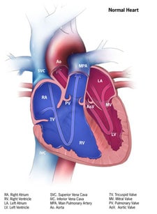
Human Heart - Anatomy, Functions and Facts about Heart The external structure of the heart has many blood vessels that form a network, with other major vessels emerging from within the structure. The blood vessels typically comprise the following: Veins supply deoxygenated blood to the heart via inferior and superior vena cava, and it eventually drains into the right atrium. A Diagram of the Heart and Its Functioning Explained in Detail Heart pumps pure blood to different parts of the body and then takes the deoxygenated blood from all the parts to the lungs for oxygenation. Normally in a minute the heart beats 72 times and pumps around 1,500 to 2,000 gallons of blood per day. Let's check out heart diagram which can help you to understand functioning of the heart in a better ... PDF Anatomy of Heart Labeled and Unlabeled Images ascending aorta pulmonary valve opening of superior vena cava right atrium fossa ovalis tricuspid valve right ventricle trabeculae carneae (a) anterior dissection of the heart 2019 pearson education, inc, pu monary trunk openings of left pulmonary veins left atrium aortic valve mitral (bicuspid) va ve chordae tendineae papillary muscle left …
Heart structure and labels. Structure of the Heart | The Franklin Institute Structure of the Heart Although most people know that the human heart doesn't bear much resemblance to a heart drawn on a Valentine's Day card, the image can still be a useful way to learn and remember the parts of the heart. The heart consists of four chambers: two atria on the top and two ventricles on the bottom. Heart Anatomy: size, location, coverings and layers ... Heart Anatomy. The heart is around the size of a fist and weighs between 250-350 grams (less than a pound). Enclosed within the mediastinum, the medial cavity of the thorax, the heart extends obliquely from the second rib to the fifth intercostal space. It rests on the superior surface of the diaphragm, lies posterior to the sternum and ... The Anatomy of the Heart, Its Structures, and Functions The heart is the organ that helps supply blood and oxygen to all parts of the body. It is divided by a partition (or septum) into two halves. The halves are, in turn, divided into four chambers. The heart is situated within the chest cavity and surrounded by a fluid-filled sac called the pericardium. This amazing muscle produces electrical ... Heart: Anatomy and Function What are the parts of the heart's anatomy? The parts of your heart are like the parts of a house. Your heart has: Walls. Chambers (rooms). Valves (doors). Blood vessels (plumbing). Electrical conduction system (electricity). Heart walls Your heart walls are the muscles that contract (squeeze) and relax to send blood throughout your body.
Diagram of Human Heart and Blood Circulation in It | New ... A heart diagram labeled will provide plenty of information about the structure of your heart, including the wall of your heart. The wall of the heart has three different layers, such as the Myocardium, the Epicardium, and the Endocardium. Here's more about these three layers. Epicardium Heart Labels - Printable or Custom Printed Stickers ... Use our free specialty shape label templates to easily personalize your heart labels online. Customize one of our free designs or upload your own graphics and then choose the printing option that works best for you. Order your blank heart labels or custom printed heart labels and stickers online and get free shipping on orders of $50 more. Heart Diagram with Labels and Detailed Explanation The heart is located under the ribcage, between the lungs and above the diaphragm. It weighs about 10.5 ounces and is cone shaped in structure. It consists of the following parts: Heart Detailed Diagram Heart - Chambers There are four chambers of the heart . The upper two chambers are the auricles and the lower two are called ventricles. Heart: illustrated anatomy - e-Anatomy - IMAIOS This interactive atlas of human heart anatomy is based on medical illustrations and cadaver photography. The user can show or hide the anatomical labels which provide a useful tool to create illustrations perfectly adapted for teaching. Anatomy of the heart: anatomical illustrations and structures, 3D model and photographs of dissection.
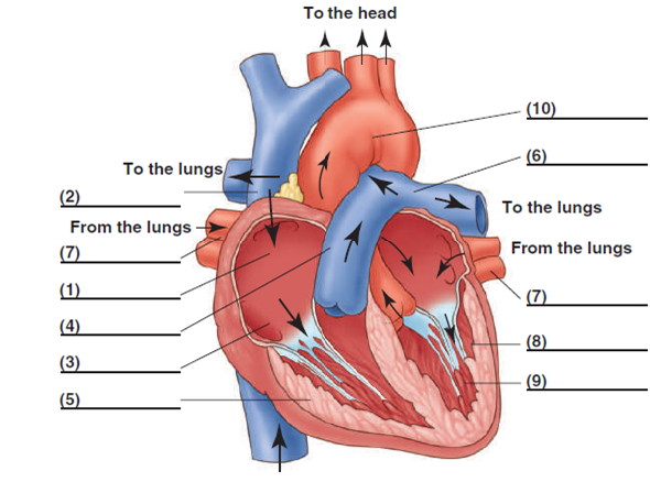
Labelling the heart — Science Learning Hub Blood transports oxygen and nutrients to the body. It is also involved in the removal of metabolic wastes. In this interactive, you can label parts of the human heart. Drag and drop the text labels onto the boxes next to the diagram. Selecting or hovering over a box will highlight each area in the diagram. Heart Labeling Quiz: How Much You Know About ... - ProProfs Here is a Heart labeling quiz for you. The human heart is a vital organ for every human. The more healthy your heart is, the longer the chances you have of surviving, so you better take care of it. Take the following quiz to know how much you know about your heart. Questions and Answers. 1. Label the heart — Science Learning Hub Label the heart Add to collection In this interactive, you can label parts of the human heart. Drag and drop the text labels onto the boxes next to the diagram. Selecting or hovering over a box will highlight each area in the diagram. Pulmonary vein Right atrium Semilunar valve Left ventricle Vena cava Right ventricle Pulmonary artery Aorta Heart Anatomy: Labeled Diagram, Structures, Blood Flow ... There are 4 chambers, labeled 1-4 on the diagram below. To help simplify things, we can convert the heart into a square. We will then divide that square into 4 different boxes which will represent the 4 chambers of the heart. The boxes are numbered to correlate with the labeled chambers on the cartoon diagram.
31 Label The Heart Answers - Labels Database 2020
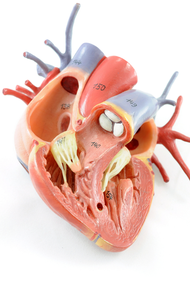
Human Heart Models | Heart Anatomy Models | Vitality Medical The heart model is available with either 2-, 4-, and 5-part magnetic sections to isolate any part of the model during the demonstration. The heart model with labels is hand-painted with vivid colors to illustrate the papillary muscles, heart valves, and adjacent structures.
Cardiovascular System - the heart Flashcards | Easy Notecards
Label Heart Anatomy Diagram Printout - EnchantedLearning.com Every day, the heart pumps about 2,000 gallons (7,600 liters) of blood, beating about 100,000 times. Label the heart anatomy diagram below using the heart glossary . Note: On the diagram, the right side of the heart appears on the left side of the picture (and vice versa) because you are looking at the heart from the front.

Pin by Daffodilcooper on BSC2086 | Heart model, Anatomy models labeled, Cardiac anatomy
Heart Diagram with Labels and Detailed Explanation - BYJUS Diagram of Heart. The human heart is the most crucial organ of the human body. It pumps blood from the heart to different parts of the body and back to the heart. The most common heart attack symptoms or warning signs are chest pain, breathlessness, nausea, sweating etc. The diagram of heart is beneficial for Class 10 and 12 and is frequently ...
How the Heart Works | Congenital Heart Defects | NCBDDD | CDC
Heart Anatomy | Anatomy and Physiology - Lumen Learning Internal Structure of the Heart. Recall that the heart's contraction cycle follows a dual pattern of circulation—the pulmonary and systemic circuits—because of the pairs of chambers that pump blood into the circulation. In order to develop a more precise understanding of cardiac function, it is first necessary to explore the internal ...

Heart Anatomy/Physiology
The structure of the heart - Structure and function of the ... It is located in the middle of the chest and slightly towards the left. The heart is a large muscular pump and is divided into two halves - the right-hand side and the left-hand side. The...

Labeled Diagram Of The Heart Simple - Photos Idea
Heart Blood Flow | Simple Anatomy Diagram, Cardiac ... The anatomy of the heart was made easy in a previous EZmed video and post, where we learned tricks to remember the main cardiac structures shown below. Check out the anatomy of the heart linked below, as that will be a great review of the main cardiac structures before learning the blood flow! Heart Anatomy: Labeled Diagram, Structures ...
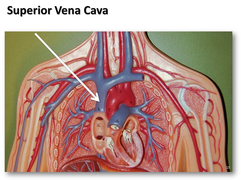
Superior vena cava - The Anatomy of the Veins Visual Guide… | Flickr
How to Draw the Internal Structure of the Heart (with ... To draw the internal structure of a human heart, follow the steps below. Part 1 Finding a Diagram 1 To find a good diagram, go to Google Images, and type in "The Internal Structure of the Human Heart". Find an image that displays the entire heart, and click on it to enlarge it. 2 Find a piece of paper and something to draw with.

heart diagram no labels
Heart Anatomy Labeling Game About this Quiz This is an online quiz called Heart Anatomy Labeling Game There is a printable worksheet available for download here so you can take the quiz with pen and paper. Your Skills & Rank Total Points 0 Get started! Today's Rank -- 0 Today 's Points One of us! Game Points 19 You need to get 100% to score the 19 points available Actions
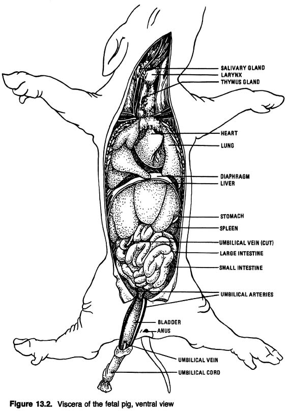
Anatomical Drawings of a Fetal Pig
Human Heart (Anatomy): Diagram, Function, Chambers ... Chambers of the Heart The heart is a muscular organ about the size of a fist, located just behind and slightly left of the breastbone. The heart pumps blood through the network of arteries and...

Medical Encyclopedia - Structure: Structure of the Heart - Aviva | THE HEART | Pinterest | Heart ...
Human Heart Diagram Labeled | Science Trends List Of Heart Structures Heart Chambers Ventricles - The bottom two heart chambers. Atra - The upper two heart chambers. Wall Of The Heart Sinoatrial Node - A collection of tissue that releases electrical impulses and defines the rate of contraction for the heart. Atrioventricular Bundle - The fibers which transmit cardiac impulses.

China U. S Trade War Heading To Economic Collapse : heading,News, breakingnews, globalnews ...
Structure of the Heart | SEER Training The human heart is a four-chambered muscular organ, shaped and sized roughly like a man's closed fist with two-thirds of the mass to the left of midline. The heart is enclosed in a pericardial sac that is lined with the parietal layers of a serous membrane. The visceral layer of the serous membrane forms the epicardium. Layers of the Heart Wall

Simple heart diagram to label by kpendlebury - Teaching Resources - TES
PDF Anatomy of Heart Labeled and Unlabeled Images ascending aorta pulmonary valve opening of superior vena cava right atrium fossa ovalis tricuspid valve right ventricle trabeculae carneae (a) anterior dissection of the heart 2019 pearson education, inc, pu monary trunk openings of left pulmonary veins left atrium aortic valve mitral (bicuspid) va ve chordae tendineae papillary muscle left …

Blood Vessels: Arteries, Capillaries & More - Video & Lesson Transcript | Study.com
A Diagram of the Heart and Its Functioning Explained in Detail Heart pumps pure blood to different parts of the body and then takes the deoxygenated blood from all the parts to the lungs for oxygenation. Normally in a minute the heart beats 72 times and pumps around 1,500 to 2,000 gallons of blood per day. Let's check out heart diagram which can help you to understand functioning of the heart in a better ...
Label Heart Structure
Human Heart - Anatomy, Functions and Facts about Heart The external structure of the heart has many blood vessels that form a network, with other major vessels emerging from within the structure. The blood vessels typically comprise the following: Veins supply deoxygenated blood to the heart via inferior and superior vena cava, and it eventually drains into the right atrium.
32 Label The Diagram Of The Heart - Labels Database 2020

Labelling the heart diagram Quiz
Post a Comment for "42 heart structure and labels"